Select a module to begin
ECG Leads, Arteries, Locations, and Clinical Pearls
12 Questions • Exhaustive ModeCorrect Answer:
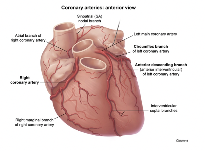
| ECG Leads | MI Location | Affected Artery |
|---|---|---|
| V1–V2 | Septal | LAD (Proximal) |
| V3–V4 | Anterior | LAD (Distal) |
| V1–V4 | Anteroseptal | LAD |
| V5–V6, I, aVL | Lateral | LCX |
| V1–V6, I, aVL | Anterolateral | LAD or LCX |
| II, III, aVF | Inferior | RCA (85%) or LCX (15%) |
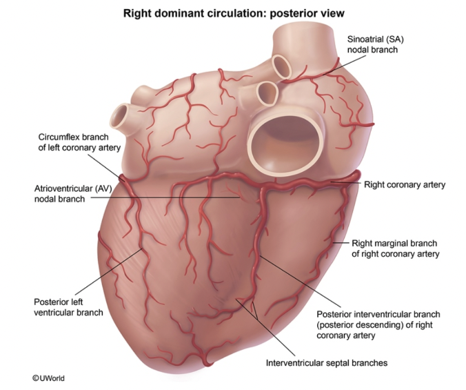
| Condition | ECG Findings | Affected Artery | Clinical Pearl |
|---|---|---|---|
| Posterior MI | ST-dep V1–V3; Tall R V1–V2 | PDA (from RCA) | Use V7–V9 to see elevation |
| Right Ventricle | ST-elev V4R | RCA (Marginal) | No Nitrates (preload dependent) |